
Cell and tissue imaging
Please find below an overview of equipment that falls under Cell and Tissue Imaging. Scroll down to find more information per equipment (e.g. contact and registration details).
| Equipment |
|---|
| Ultrawide ‘field-of-view’ multi-foton-microscope |
| JPK NanoWizard 4 ultraspeed |
| Metafer 5 microscope and image analysing system |
| Light sheet |
| STED microscope |
| Leica Bond RX IHC and ISH Research Platform |
| Incucyte SX5 HD/3CLR |
Ultrawide ‘field-of-view’ multi-foton-microscope
Location: Ee-739
Registration: no open registration. Please contact Miao Ping Chien to discuss your experiment. This machine can only be used by someone from Miao’s group.
Contact person:
Miao Ping Chien
E: m.p.chien@erasmusmc.nl
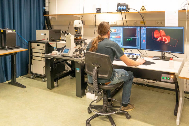
UFO2 is a custom-built high-throughput single cell screening microscope that allows one to screen, identify and isolate aggressive or abnormal tumorigenic cancer cells followed by downstream single cell genomic, transcriptomic or proteomic profiling. These profiles can then be used for target gene discovery for biomarker or drug development.
JPK NanoWizard 4 ultraspeed
Location: Ee-631a
Registration: please contact Dejan Ristic
Contact person:
Dejan Ristic
E: d.ristic@erasmusmc.nl
This instrument is used for high speed image analysis of molecular processes, such as the dynamics of DNA repair proteins after DNA damage.
Metafer 5 microscope and image analysing system
Location: Ee-775
Registration: follow this link
Contact person:
Arnab Ray Chaudhuri
E: a.raychaudhuri@erasmusmc.nl
The Metafer 5 and associated robotic sample feeder is a upright fluorescent microscope ( 4 color filters), specialized in the automated detection and imaging of metaphase spreads. It is also capable of detecting and scoring micronuclei and imaging of comet assays (using the trevigen system for which adapters are available.) Finally, it can also be use for immunofluorescence analysis of fixed cells only in slides (used for scanning only and requires other specialized softwares for quantitative analysis). Objectives available: 20X air, 40X air, 60X oil.
Light Sheet

Location: Ee-1315
Registration: follow this link
Registration for first time users: follow this link
Website: follow this link
Contact person:
Gert van Cappellen (registration)
E: w.vancappellen@erasmusmc.nl
Martijn de Gruiter (training)
E: h.degruiter@erasmusmc.nl
With a lighsheet mciroscope it’s possible to make very fast 3D images from small organs or organisms. The technique will be used directly for study in zebrafish brain and tumor development. In the near future, it will become more and more important for organoid studies and we want to research the possibility to directly study human biopsies, especially needle biopsies from different natures (diseases).
STED microscope
Location: Be-311
Registration: follow this link
Registration for first time users: follow this link
Website: follow this link
Contact person:
Gert van Cappellen (registrations)
E: w.vancappellen@erasmusmc.nl
Martijn de Gruiter (training)
E: h.degruiter@erasmusmc.n
The Leica STED (STimulated Emission Depletion) is a super-resolution microscope developed by the Nobel prize laureate Stefan Hell. With this microscope we are able to fill the gap between the Electron Microscope and the Confocal Microscope. It is now possible to study on a sub-cellular level what is going wrong during diseases. Especially effect of mutations in proteins, which are often the cause of diseases can now be studied in much future detail.
Leica Bond RX IHC and ISH Research Platform
Location: Be-454
Registration: follow this link
Website: follow this link
Contact person:
Cor Berrevoets
E: c.berrevoets@erasmusmc.nl
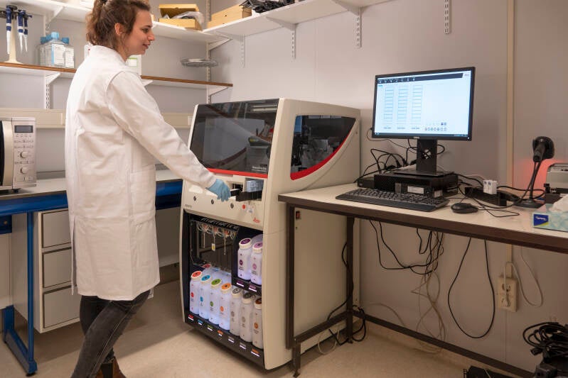
Leica BOND RX is a fully automated stainer that enables high-throughput processing of tumor sections, and is compatible with a wide array of staining platforms (i.e., immune fluorescence, immune histochemistry, in situ hybridization, multiplex stainings etc.). With Leica all technical steps become customized, such as preparation (baking and dewaxing), antigen retrieval (incubation time and temperature), adding reagents (probe or antibody application), as well as staining conditions (incubation time, temperature, dispense type). Also, Leica enables execution of different protocols in an automated fashion to maximize assay viability in minimal time, and thanks to small volumes used, with minimal reagents. This equipment is central towards the automation of imaging of tumor tissues with Vectra 3.0, thereby reducing costs and variability and increasing viability and throughput.
Incucyte SX5 HD/3CLR
Location: Be-460
Registration: please contact Cor Berrevoets
Contact person:
Cor Berrevoets
E: c.berrevoets@erasmusmc.nl
The Incucyte SX5 HD/3CLR is a real-time quantitative live-cell imaging and analysis system to visualise and quantify cell behavior over time. The Incucyte automatically gathers and analyzes cellular images with 3 different fluorescent channels (green, orange and near-infrared) within a standard laboratory incubator, and creates time-lapsed, kinetic profiles from living cancer cells or immune cells over days and weeks. With these measurements our understanding of active biological processes or interactions (and their targeting) involving tumor cells substantially increases.